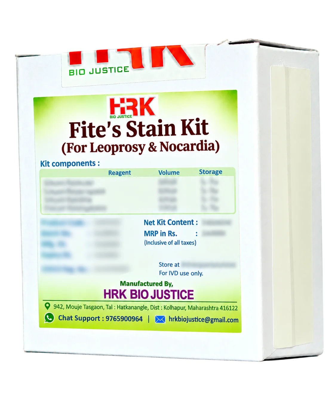
Fite’s Stain Kit

Product images are for reference only and may vary from the actual product.

Fite’s Stain Kit

For Leprosy & Nocardia

In Stock

Share Now :
Fite’s Stain is a modified acid-fast staining method designed for the histological demonstration of Mycobacterium leprae and Nocardia in paraffin-embedded tissue sections. The method uses an oil-containing deparaffinization step to help preserve the relatively delicate acid-fastness of M. leprae.
  • Special stain for Mycobacterium leprae and Nocardia
  • Suitable for formalin-fixed, paraffin-embedded tissue sections
  • Modified acid-fast technique using xylene–peanut oil deparaffinization
  • Helps preserve the acid-fast characteristics of delicate organisms
  • Carbol fuchsin provides strong red/magenta staining of acid-fast organisms
  • Methylene blue provides a contrasting blue background
  • Suitable for routine histopathology laboratories
  • Can be used for both Leprosy and Nocardia staining protocols, with appropriate differentiation conditions.
  • The waxy, lipid-rich cell wall of acid-fast organisms retains carbol fuchsin despite controlled decolorization. In Fite staining, xylene–peanut oil is used for deparaffinization to reduce loss of acid-fastness of M. leprae. After staining with carbol fuchsin, controlled differentiation removes stain from the surrounding tissue while acid-fast organisms remain red. Methylene blue provides the contrasting background.
  • Typical result:
    1. 1. Leprosy bacilli / acid-fast organisms: Bright red to magenta
      2. Nuclei/background: Blue
  • Optimized for delicate acid-fast organisms: Particularly useful for demonstrating M. leprae, whose acid-fastness can be compromised by conventional deparaffinization.
  • Clear organism-to-background contrast: Red/magenta bacilli against a blue background facilitates microscopic recognition.
  • Useful in tissue diagnosis: Supports histopathological evaluation of suspected leprosy and Nocardia infection.
  • Paraffin-section compatible: Designed for routine FFPE histology specimens.
  • Convenient laboratory workflow: Ready-to-use kit formats can provide standardized staining reagents and reduce individual reagent preparation.
  • Suitable for microscopic confirmation: Organisms can be examined at high magnification, including oil-immersion microscopy when required.

Related products

Recently Viewed Products

Scroll to Top

Where Histology meets certainty ….!

Office Address

849, Mouje Tasgaon,
Tal. Hatkanangale,
Dist. Kolhapur - 416122
Maharashtra, India.

phone number

+91 927 033 6969