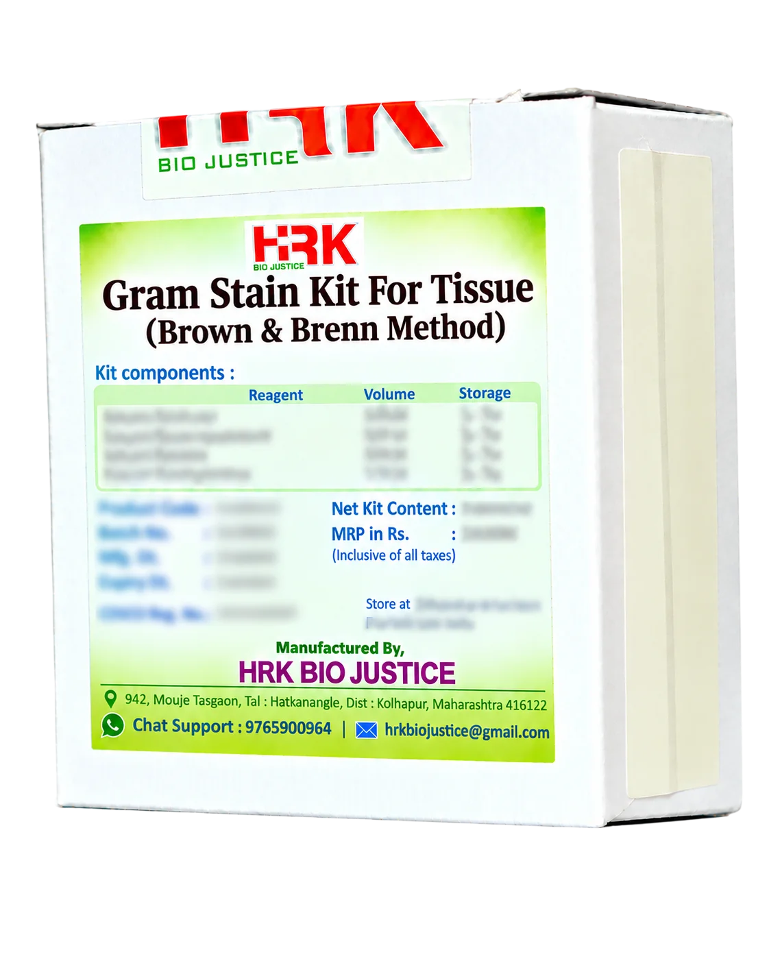
Gram Stain Kit for Tissue

Product images are for reference only and may vary from the actual product.

Gram Stain Kit for Tissue

Brown & Brenn Method

In Stock

Share Now :
  • Gram Stain Kit for Tissue – Brown & Brenn Method is a modified Gram staining system designed for the demonstration and differentiation of Gram-positive and Gram-negative bacteria in formalin-fixed, paraffin-embedded tissue sections.
  • The method provides contrasting staining of bacterial organisms against the surrounding tissue background, making it useful for detecting bacterial colonies and assessing their distribution and morphology in histopathological specimens.
  • Modified Brown & Brenn Gram staining method for tissue sections
  • Specifically designed for histopathological specimens
  • Suitable for formalin-fixed, paraffin-embedded tissue sections
  • Demonstrates both Gram-positive and Gram-negative bacteria
  • Provides strong colour contrast between bacteria and tissue components
  • Gram-positive organisms typically stain blue to purple
  • Gram-negative organisms typically stain red to pink
  • Counterstaining enhances tissue morphology and background contrast
  • Useful for bacterial localization within tissue sections
  • Suitable for routine histopathology and special-stain applications
  • Designed for microscopic examination of stained tissue sections
  • The Brown & Brenn method is a modified Gram staining technique adapted for tissue sections. Crystal violet and iodine form a dye–iodine complex within bacterial cells. During differentiation, Gram-positive organisms retain the primary stain more effectively because of their thick peptidoglycan cell wall, whereas Gram-negative organisms lose the primary complex more readily.
  • A contrasting counterstain subsequently colours Gram-negative organisms and selected tissue components, producing a differential colour reaction. The Brown & Brenn modification incorporates additional staining components to improve the visualization of bacteria within tissue sections while maintaining useful tissue-background contrast.
  • Effective Bacterial Demonstration
    1. Provides a practical histochemical method for demonstrating bacteria in tissue sections.
  • Differentiation of Gram Reaction
    1. Allows morphological differentiation between Gram-positive and Gram-negative organisms based on their characteristic staining reactions.
  • Enhanced Tissue Contrast
    1. Provides contrasting bacterial and tissue staining, facilitating microscopic localization of microorganisms.
  • Useful for Tissue-Associated Infections
    1. Can assist in demonstrating bacteria associated with inflammatory, necrotic or infected tissue areas.
  • Improved Bacterial Localization
    1. Helps reveal the distribution and relationship of bacterial organisms within the tissue architecture.
  • Suitable for Paraffin Sections
    1. Designed for routine formalin-fixed, paraffin-embedded histopathological sections.
  • Supports Histopathological Assessment
    1. Adds microbiological information to routine tissue morphology and inflammatory findings.
  • Useful as an Adjunctive Special Stain
    1. Can complement other microbiological stains and ancillary investigations when bacterial infection is suspected.
  • Typical Applications :
    1. 1. Demonstration of bacteria in tissue sections
      2. Gram-positive and Gram-negative bacterial differentiation
      3. Histopathological evaluation of suspected bacterial infections
      4. Detection of bacterial colonies in tissue
      5. Examination of infected and inflammatory lesions
      6. Localization of bacteria within tissue architecture
      7. Routine histopathology special staining
      8. Paraffin-embedded tissue sections.

Related products

Recently Viewed Products

Scroll to Top

Where Histology meets certainty ….!

Office Address

849, Mouje Tasgaon,
Tal. Hatkanangale,
Dist. Kolhapur - 416122
Maharashtra, India.

phone number

+91 927 033 6969