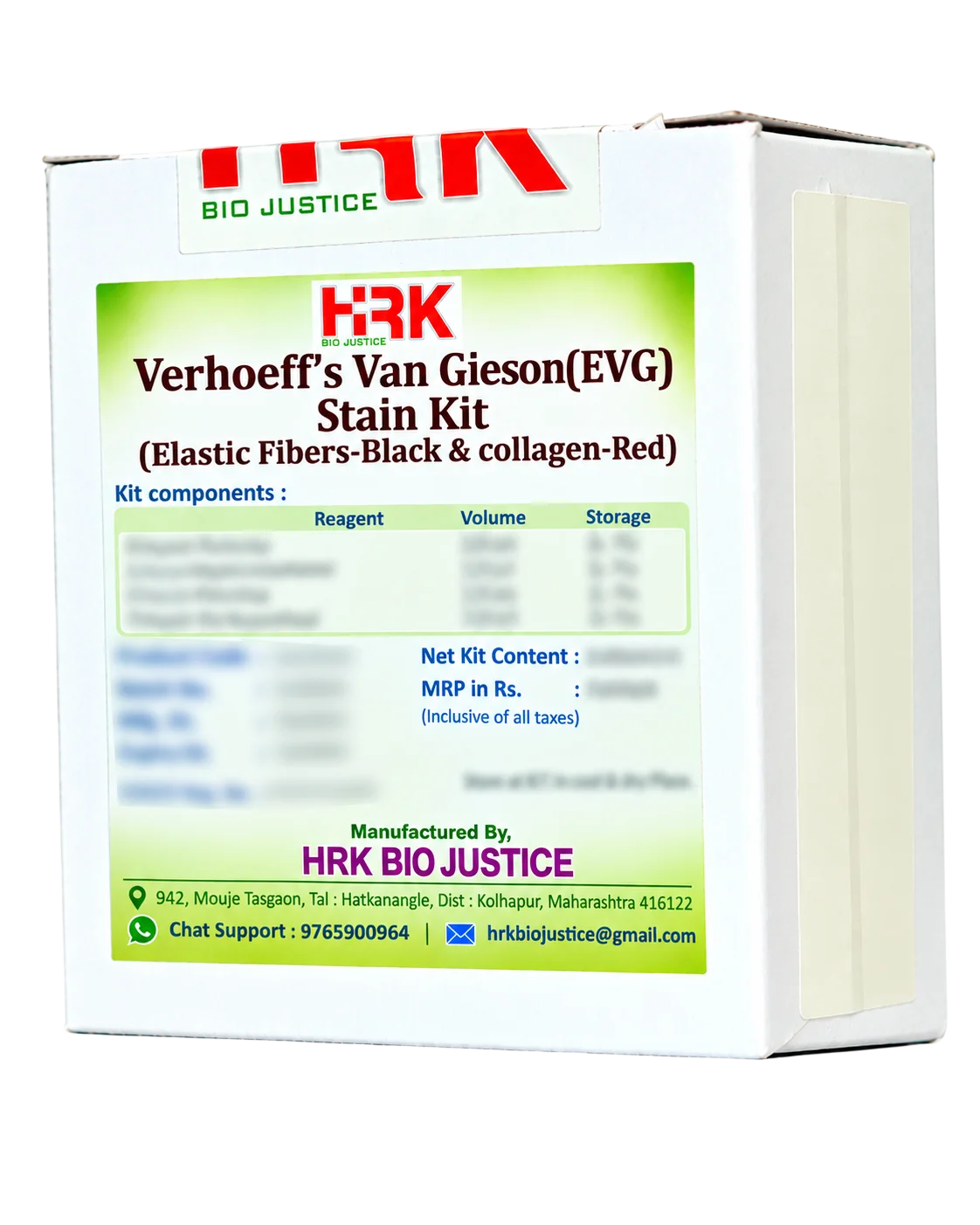
Verhoeff’s Van Gieson (EVG) Stain Kit

Product images are for reference only and may vary from the actual product.

Verhoeff’s Van Gieson (EVG) Stain Kit

Elastic Fibers-Black & Collagen-Red

In Stock

Share Now :
  • Verhoeff’s Van Gieson (EVG) Stain Kit is a special histopathology staining method for the demonstration of elastic fibres and collagen fibres in tissue sections. The Verhoeff elastic stain selectively demonstrates elastic fibres in black, while the Van Gieson counterstain provides a contrasting red colour to collagen.
  • The stain is widely used for evaluating connective-tissue architecture, vascular elastic laminae and alterations in elastic fibre distribution.
  • Clear demonstration of elastic fibres in black
  • Collagen fibres stained red with Van Gieson counterstain
  • Provides strong colour contrast for microscopic evaluation
  • Useful for demonstrating elastic laminae in blood vessels
  • Suitable for routine histopathology and special-stain applications
  • Applicable to a wide range of tissue sections
  • Convenient kit format for standardized manual staining
  • Supports consistent staining when used according to the recommended protocol
The Verhoeff component uses a hematoxylin-based iron–hematoxylin staining system containing ferric ions and iodine/iodide. Elastic fibres have a strong affinity for the Verhoeff stain and retain the dye complex after controlled differentiation, appearing black.
The subsequent Van Gieson counterstain provides a contrasting background: collagen appears red, while other tissue elements take up contrasting yellow tones. This combination makes the elastic fibre network readily distinguishable from collagen and surrounding tissue.

    1. Applications

  • Demonstration of elastic fibres in arteries and other blood vessels
  • Evaluation of elastic laminae
  • Lung and respiratory tissue architecture
  • Skin and connective tissue
  • Aorta and large-vessel specimens
  • Assessment of elastic fibre alterations in pathological tissue
  • Histopathology teaching and research
  • High-contrast visualization: Black elastic fibres against a contrasting red collagen background facilitate microscopic identification.
  • Excellent architectural detail: Helps assess the relationship between elastic fibres, collagen and surrounding tissue structures.
  • Vascular evaluation: Particularly useful for demonstrating internal and external elastic laminae in blood vessels.
  • Dual-purpose special stain: Demonstrates two important connective-tissue components within the same section.
  • Convenient workflow: Provides the essential staining reagents in a practical kit format.
  • Broad tissue application: Suitable for vascular, pulmonary, skin and other connective-tissue specimens.
  • Consistent staining: Supports reproducible results when standardized staining and differentiation conditions are maintained.

Related products

Recently Viewed Products

Scroll to Top

Where Histology meets certainty ….!

Office Address

849, Mouje Tasgaon,
Tal. Hatkanangale,
Dist. Kolhapur - 416122
Maharashtra, India.

phone number

+91 927 033 6969