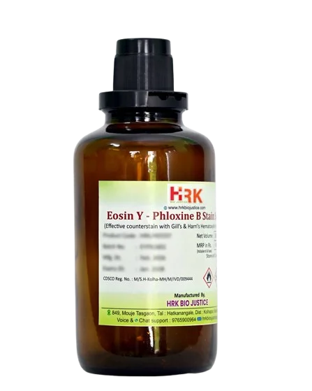
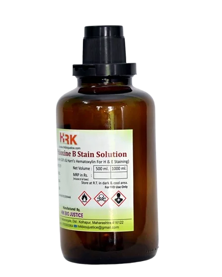
Eosin Y-Phloxine B Stain Solution

Product images are for reference only and may vary from the actual product.

Eosin Y-Phloxine B Stain Solution

Bright Red Variant for High Contrast H&E

In Stock

Share Now :
Eosin Y- Phloxine B is a premium dual-dye formulation where Phloxine B (a brilliant red xanthene dye) is added to Eosin Y. While standard eosin gives a standard pink, Phloxine B imparts an intense, fiery red-orange contrast that makes cytoplasm, RBCs, and collagen pop under the microscope. Preferred for high-cellular biopsies, oncology specimens, and digital slide scanning.
  • Dual-Dye Technology: Combination of Eosin Y (Yellowish) + Phloxine B (Bluish-Red) for extended color spectrum
  • Brilliant Red Shade: Gives 3-Tone Eosinophilia – Pink, Orange-Red & Fiery Red vs Single Pink in routine eosin
  • Phloxine B Enhanced: 5-10% Phloxine B w.r.t Eosin – Scientifically optimized ratio
  • Alcoholic Base: 70-90% Ethanol/Methanol formulation – Rapid penetration, no overstaining
  • High-Contrast Formulation: Specifically for WSI / Digital Pathology scanners
  • Ready-to-Use 1% w/v: No dilution, no filtering required
  • ISO 13485 Manufactured: Batch to batch consistency for NABL labs
Bright Red Variant works on differential eosinophilia principle:

  • Eosin Y – Stains general cytoplasm and muscle in light pink-orange.
  • Phloxine B – Being more acidic and larger molecule, it selectively binds to highly eosinophilic structures with high affinity.
  • Result – 3 Color Contrast:
    1. General Cytoplasm: Light Pink
      Collagen / Muscle / Keratin: Deep Red-Orange
      RBCs / Eosinophils / Paneth Cell Granules: Fiery Brilliant Red
    This differential staining makes it easy to differentiate 3 tissues in one field, which is impossible with standard single-dye eosin.

  • For Pathologist:
    1. Better Diagnosis in Cancer: Tumor keratin, necrosis, and cell borders are much clearer in SCC, Breast, Cervix.
      Easy Hemorrhage Detection: RBCs glow, you won’t miss vascular invasion.
      No Confusion: Muscle vs Collagen vs Keratin – all different reds.
  • For Lab Owner:
    1. Wins Digital Pathology: Scanned slides look stunning on monitor. Perfect for Morphle, Leica, Philips scanners and AI.
      Saves Special Stain Cost: In many liver / skin biopsies, you can avoid Trichrome just to see collagen.
      30% Faster: Stains in 30 sec. Increases daily throughput.
  • For Teaching Labs:
    1. Students Learn Faster: 3 colors are easier to teach than 1 pink.
      Projector Ready: Even from last bench, slide looks contrasty.
  • Technical Specifications :
  • Specification Details
    Composition Eosin Y + Phloxine B in Alcoholic Solution
    pH 4.8 ± 0.2 (With Glacial Acetic Acid)
    Color Clear Dark Reddish-Orange Solution
    Staining Time 30 sec – 90 sec
    Compatibility All Hematoxylins (Harris, Mayer, Gill)
    Shelf Life 24 Months
    Storage 15-30°C
    Pack Size 500ml, 1L, 5L

Related products

Recently Viewed Products

Scroll to Top

Where Histology meets certainty ….!

Office Address

849, Mouje Tasgaon,
Tal. Hatkanangale,
Dist. Kolhapur - 416122
Maharashtra, India.

phone number

+91 927 033 6969