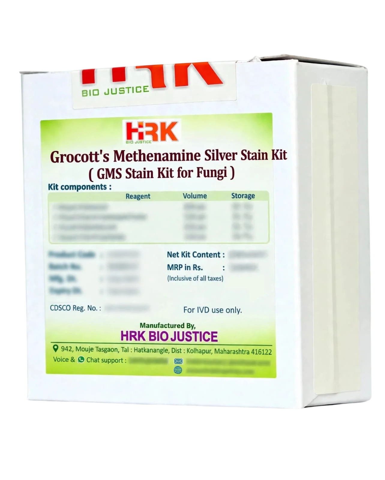
Grocott’s Methenamine Silver Stain Kit
GMS Stain Kit for Fungi
In Stock
SKU
N/A
Categories : Digital-Optimized H&E & Special Stains, IHC Compatible Histopathology Reagents (Fix to Mount)
Share Now :
Grocott’s Methenamine Silver (GMS) Stain Kit is a special histological staining system designed for the demonstration and screening of fungal organisms in tissue sections and cytological preparations. It is particularly useful for highlighting fungal cell walls, including yeast and hyphal forms, against a contrasting tissue background. GMS is widely used for detecting fungal elements and Pneumocystis organisms in histopathology.
- Special stain for fungal organisms in histological sections
- Produces black to dark-brown fungal elements against a contrasting background
- Demonstrates yeast, hyphae and other fungal structures
- Useful for screening invasive and opportunistic fungal infections
- Suitable for formalin-fixed, paraffin-embedded (FFPE) tissue sections
- Useful for demonstrating Pneumocystis organisms
- Provides strong contrast for detection of small or sparse fungal elements
- Complete staining system designed for routine histopathology laboratories
- Suitable for use alongside H&E and PAS for complementary evaluation
GMS staining is based on the oxidation of polysaccharides in fungal cell walls followed by reduction of silver ions to metallic silver. Chromic acid oxidizes fungal cell-wall polysaccharides to form aldehyde groups. Under alkaline conditions, the methenamine-silver complex reacts at these sites, producing black to dark-brown silver deposits that sharply outline fungal elements. A light-green counterstain provides contrast with the surrounding tissue.

- Excellent fungal visibility: Strong silver impregnation makes fungal cell walls readily identifiable.
- High contrast: Black/dark fungal structures against a green background facilitate microscopic detection.
- Sensitive screening method: GMS is particularly useful for detecting fungal elements that may be inconspicuous on routine H&E.
- Broad fungal applicability: Useful for demonstrating a wide range of fungal morphologies, including yeast and hyphal forms.
- Useful in difficult specimens: Helps reveal fungal elements within inflammatory, necrotic or granulomatous tissue.
- Diagnostic workflow support: Can be used with H&E, PAS and other ancillary stains to improve evaluation of suspected fungal infection.
- Routine laboratory application: Designed for practical use in histopathology and surgical pathology laboratories.
- Typical staining result: Fungal elements – Black to dark brown | Background – Light green.
Related products
Related products
-
IHC Compatible Histopathology Reagents (Fix to Mount)
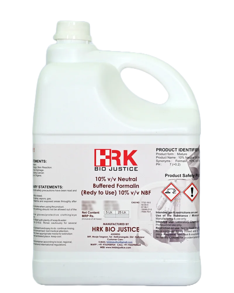
10% (v/v) Neutral Buffered Formalin
₹500.00 – ₹800.00Price range: ₹500.00 through ₹800.00 Select options This product has multiple variants. The options may be chosen on the product page -
Bone Marrow Decalcifiers-IHC Compatible RTU
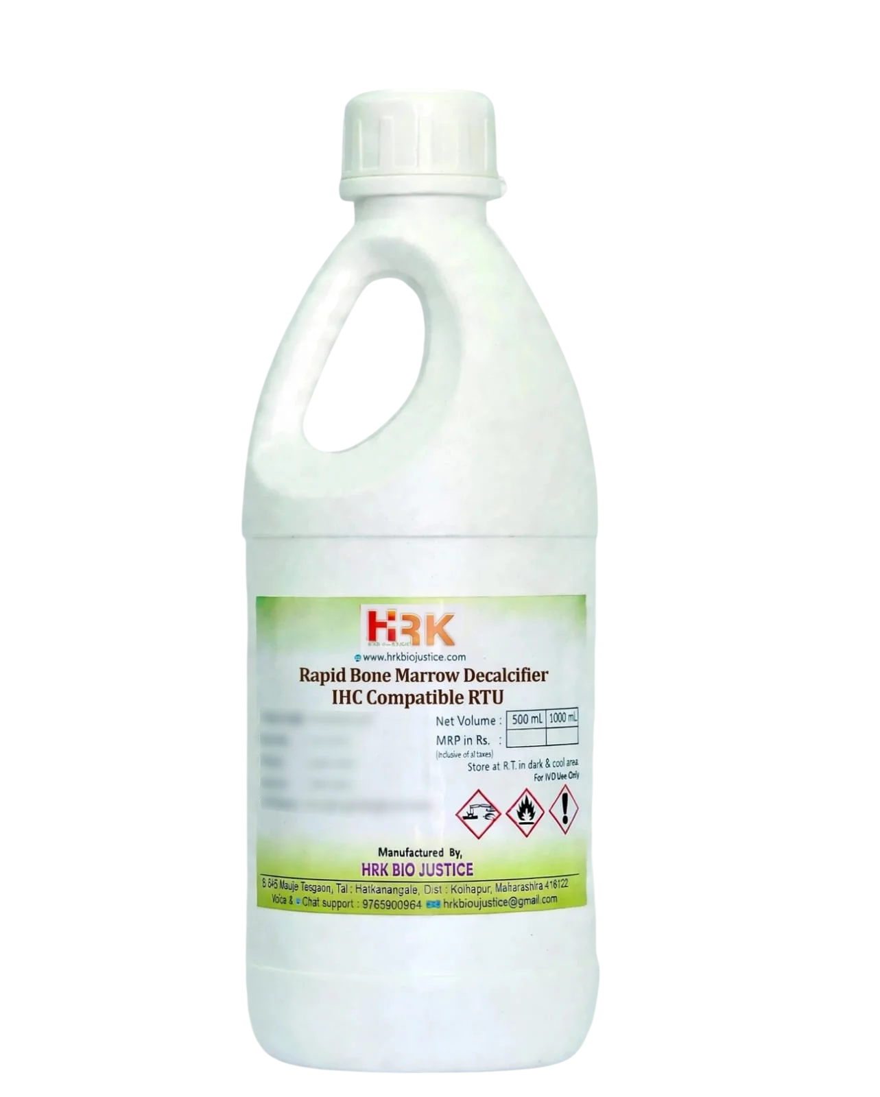
Rapid Bone Marrow Decalcifier
₹100.00 – ₹300.00Price range: ₹100.00 through ₹300.00 Select options This product has multiple variants. The options may be chosen on the product page -
Digital-Optimized H&E & Special Stains
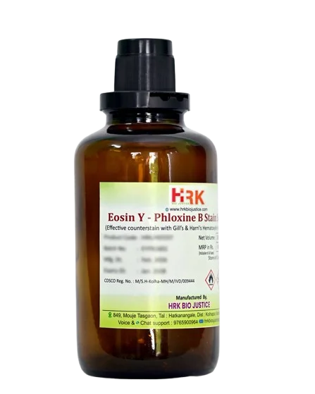
Eosin Y-Phloxine B Stain Solution
₹740.00 – ₹1,020.00Price range: ₹740.00 through ₹1,020.00 Select options This product has multiple variants. The options may be chosen on the product page
Recently Viewed Products
-
Engineered Tissue Adhesives for IHC/ ISH/ FISH
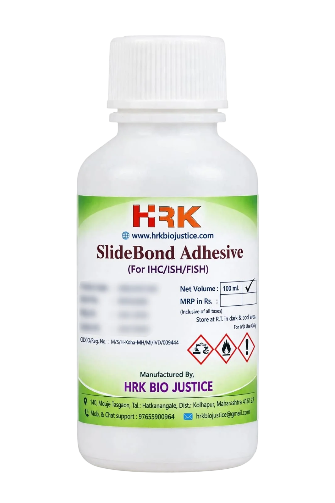
SlideBond TM Adhesive
₹535.00 – ₹1,050.00Price range: ₹535.00 through ₹1,050.00 Select options This product has multiple variants. The options may be chosen on the product page