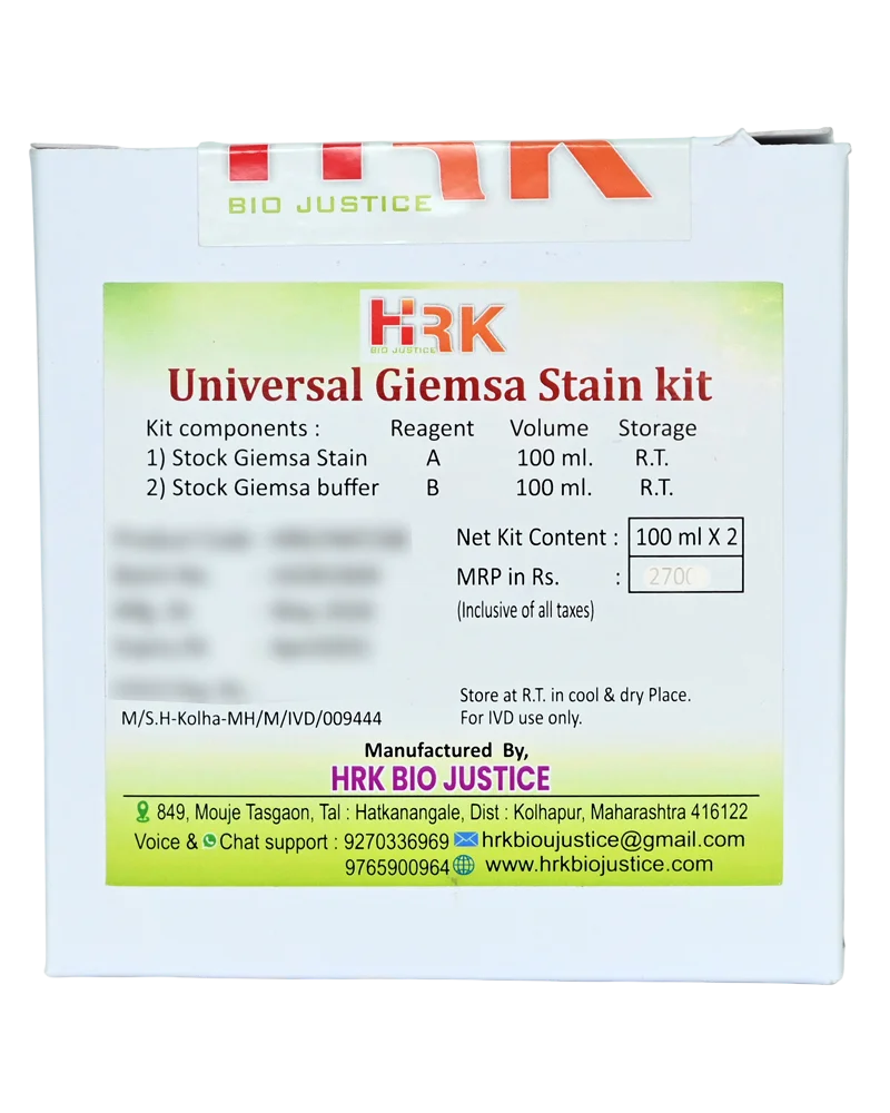
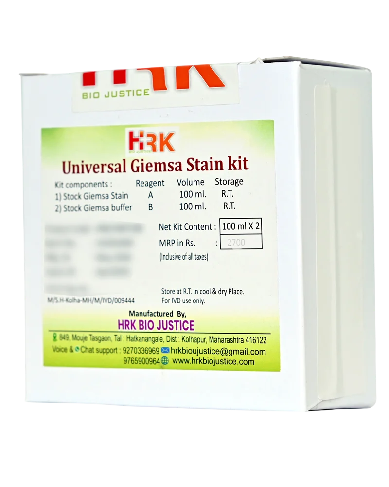
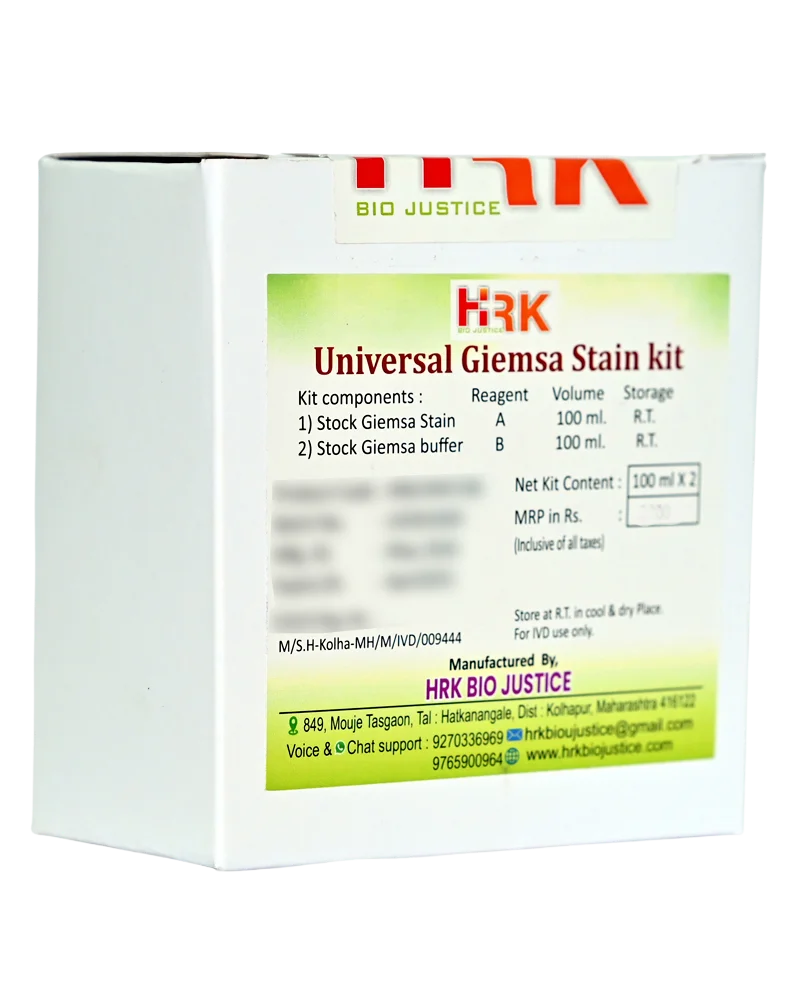
Universal Giemsa Stain Kit

Product images are for reference only and may vary from the actual product.

Universal Giemsa Stain Kit

For Hematology, Histopathology & Cytology

In Stock

Share Now :
  • Universal Giemsa Stain Kit is a versatile Romanowsky-type staining system designed for differential staining of cellular and tissue components across hematology, histopathology and cytology applications.
  • It provides characteristic blue, violet and pink staining patterns that enhance nuclear, cytoplasmic and cellular detail, supporting routine microscopic examination of appropriately prepared specimens.
  • Versatile Giemsa staining system for multiple laboratory applications
  • Suitable for hematology, histopathology and cytology workflows
  • Provides characteristic Romanowsky-type staining
  • Produces clear nuclear and cytoplasmic differentiation
  • Enhances visualization of cellular morphology
  • Suitable for peripheral blood and other appropriate cytological preparations
  • Can be used for selected tissue sections following a validated laboratory protocol
  • Supports visualization of cellular inclusions and microorganisms where appropriate
  • Convenient staining procedure
  • Designed for consistent and reproducible staining
  • Suitable for routine microscopic examination
  • Giemsa staining is based on the Romanowsky staining principle, utilizing a combination of basic and acidic dye components.
  • Basic dye components interact predominantly with acidic cellular structures such as nuclear chromatin, while acidic dye components stain basic cytoplasmic components. The interaction between these dyes produces characteristic shades ranging from blue and blue-purple to pink and violet, depending on the cellular structure and staining conditions.
  • This differential staining enhances contrast between nuclei, cytoplasm, granules and other cellular structures, facilitating morphological examination under the microscope.
  • Multi-Application Use – Suitable for appropriately validated applications across hematology, histopathology and cytology.
  • Clear Nuclear Detail – Provides characteristic staining of nuclear chromatin for morphological evaluation.
  • Distinct Cellular Differentiation – Enhances contrast between nuclear and cytoplasmic components.
  • Good Morphological Contrast – Helps visualize cellular structures and selected inclusions or microorganisms.
  • Versatile Laboratory Workflow – Reduces the need to maintain separate Giemsa staining systems for different routine applications where the same formulation is validated.
  • Consistent Staining Performance – Supports reproducible results when standardized staining conditions are maintained.
  • Convenient Routine Use – Designed for practical implementation in routine microscopy laboratories.
  • Broad Specimen Compatibility – Can be applied to suitable blood films, cytological preparations and selected tissue sections according to validated procedures.
  • Applications :
  • Application Hematology Histopathology Cytology Microscopy
    Description •Peripheral blood smears
    •Blood-cell morphology
    •Differential leukocyte examination
    •Bone marrow smears
    •Selected tissue sections requiring
    Giemsa staining
    •Demonstration of suitable cellular or microbial structures according to the laboratory’s validated protocol
    •Conventional cytology smears
    •Fine-needle aspiration preparations
    •Selected cellular specimens requiring Giemsa-type differential staining
    Suitable for morphological evaluation of appropriately prepared and fixed specimens using established laboratory procedures.

Related products

Recently Viewed Products

Scroll to Top

Where Histology meets certainty ….!

Office Address

849, Mouje Tasgaon,
Tal. Hatkanangale,
Dist. Kolhapur - 416122
Maharashtra, India.

phone number

+91 927 033 6969